Makindo Medical Notes.com |
|
|---|---|
| Download all this content in the Apps now Android App and Apple iPhone/Pad App | |
| MEDICAL DISCLAIMER:The contents are under continuing development and improvements and despite all efforts may contain errors of omission or fact. This is not to be used for the assessment, diagnosis or management of patients. It should not be regarded as medical advice by healthcare workers or laypeople. It is for educational purposes only. Please adhere to your local protocols. Use the BNF for drug information. If you are unwell please seek urgent healthcare advice. If you do not accept this then please do not use the website. Makindo Ltd | |
ECG - Bundle Branch Blocks
-
| About | Anaesthetics and Critical Care | Anatomy | Biochemistry | Cardiology | Clinical Cases | CompSci | Crib | Dermatology | Differentials | Drugs | ENT | Electrocardiogram | Embryology | Emergency Medicine | Endocrinology | Ethics | Foundation Doctors | Gastroenterology | General Information | General Practice | Genetics | Geriatric Medicine | Guidelines | Haematology | Hepatology | Immunology | Infectious Diseases | Infographic | Investigations | Lists | Microbiology | Miscellaneous | Nephrology | Neuroanatomy | Neurology | Nutrition | OSCE | Obstetrics Gynaecology | Oncology | Ophthalmology | Oral Medicine and Dentistry | Paediatrics | Palliative | Pathology | Pharmacology | Physiology | Procedures | Psychiatry | Radiology | Respiratory | Resuscitation | Rheumatology | Statistics and Research | Stroke | Surgery | Toxicology | Trauma and Orthopaedics | Twitter | Urology
Related Subjects: |ECG Basics |ECG Axis |ECG Analysis |ECG LAD |ECG RAD |ECG Low voltage |ECG Pathological Q waves |ECG ST/T wave changes |ECG LBBB |ECG RBBB |ECG short PR |ECG Heart Block |ECG Asystole and P wave asystole |ECG QRS complex |ECG ST segment |ECG: QT interval |ECG: LVH |ECG RVH |ECG: Bundle branch blocks |ECG Dominant R wave in V1 |ECG Acute Coronary Syndrome |ECG Narrow complex tachycardia |ECG Ventricular fibrillation |ECG Regular Broad complex tachycardia |ECG Crib sheets
Bundle Branch Blocks
Inspect QRS complexes for bundle branch block or fascicular block The normal QRS interval is 0.12 seconds (3 mm or 3 small squares) on the ECG. To correctly determine the QRS interval, use the lead with the widest QRS complex. If the QRS complex is less than or equal to 0.12 seconds, then no further analysis is necessary. If it is greater than 0.12 seconds, then you should try to determine the reason for the abnormally long QRS interval.
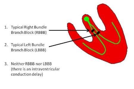
A simple approach is to consider the following three possible causes for QRS widening:

The type of bundle branch block can usually be determined from the examination of three key leads: I, V1 and V6.
Inspect QRS complexes for bundle branch block or fascicular block
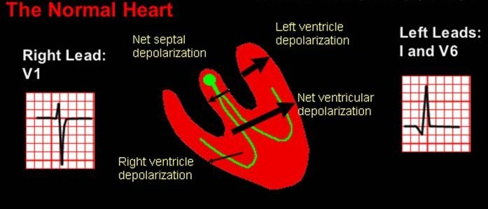
In the normal heart, at the beginning of ventricular depolarization, the QRS axis is oriented to the right because of the left-to-right depolarization of the septum. This produces a small R wave in lead V1. Immediately following septal depolarization, the left and right ventricles depolarize. The size of the left ventricle results in a predominantly leftward axis for the remainder of the QRS complex.

In the setting of RBBB, the initial part of the ECG is unchanged because septal depolarization and depolarization of the left ventricle are unaffected. However, the right ventricle depolarizes in a delayed and slow fashion. This results in a widening of the terminal part of the QRS complex and orientation of the axis of the terminal part of the QRS complex to the right.
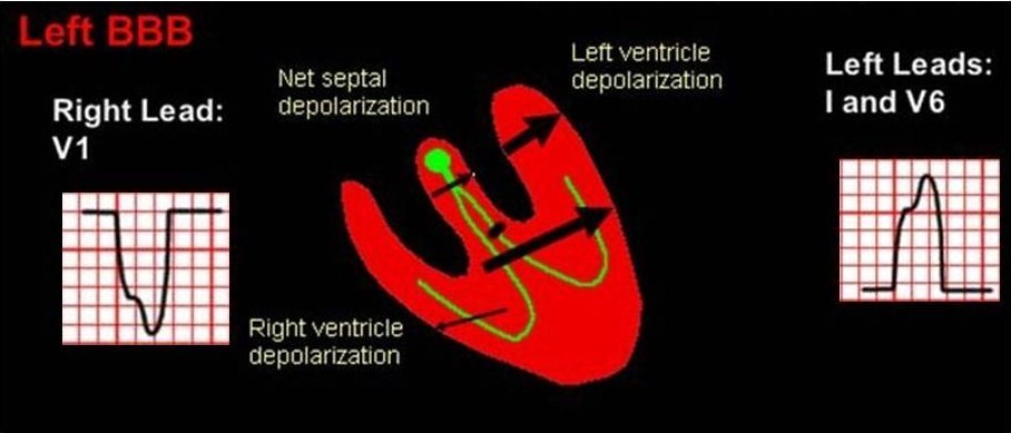
In the setting of LBBB, the septum is activated in a right to left direction, and then there is depolarization of the right and left ventricles through the right bundle. The result is that the QRS axis has a predominantly left orientation throughout and is wide secondary to the slow activation of the left ventricle.

If the ECG cannot be characterized as a typical RBBB or a typical LBBB, then it can be categorized as an intraventricular conduction delay. This will not be addressed in any more detail at this time.