Related Subjects:
|Olfactory Nerve
|Optic Nerve
|Oculomotor Nerve
|Trochlear Nerve
|Trigeminal Nerve
|Abducent Nerve
|Facial Nerve
|Vestibulocochlear Nerve
|Glossopharyngeal Nerve
|Vagus Nerve
|Accessory Nerve
|Hypoglossal Nerve
Anatomy
The facial nerve originates in the brainstem and follows a complex course through the skull to reach its target areas.
- Origin:
- Arises from the pons in the brainstem at the pontomedullary junction.
- Consists of a larger motor root and a smaller sensory root (nervus intermedius).
- Intracranial Course:
- Enters the internal acoustic meatus along with the vestibulocochlear nerve (CN VIII).
- Travels through the facial canal in the temporal bone.
- Forms the geniculate ganglion, where the sensory fibers have their cell bodies.
- Extracranial Course:
- Exits the skull through the stylomastoid foramen.
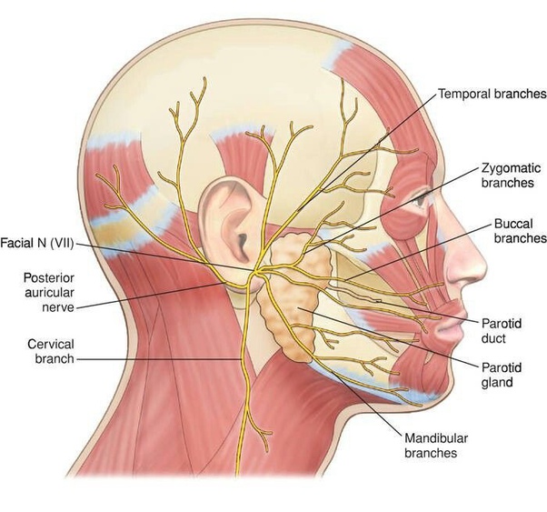
- Enters the parotid gland and divides into five major branches.
Branches of the Facial Nerve
The facial nerve has several branches that innervate various structures.
- Intracranial Branches:
- Greater Petrosal Nerve: Provides parasympathetic fibers to the lacrimal gland and mucous membranes of the nasal cavity and palate.
- Nerve to Stapedius: Innervates the stapedius muscle in the middle ear.
- Chorda Tympani: Carries taste sensations from the anterior two-thirds of the tongue and provides parasympathetic fibers to the submandibular and sublingual glands.
- Extracranial Branches:
- Temporal Branch: Innervates the muscles of the forehead and upper eyelid.
- Zygomatic Branch: Innervates the muscles around the eyes, including the orbicularis oculi.
- Buccal Branch: Innervates the muscles of the upper lip and cheek.
- Marginal Mandibular Branch: Innervates the muscles of the lower lip and chin.
- Cervical Branch: Innervates the platysma muscle in the neck.
Functions of the Facial Nerve
- Motor Functions:
- Controls the muscles of facial expression.
- Innervates the stapedius muscle in the ear, which helps dampen loud sounds.
- Sensory Functions:
- Provides taste sensations from the anterior two-thirds of the tongue via the chorda tympani.
- Parasympathetic Functions:
- Supplies parasympathetic fibers to the lacrimal glands, submandibular and sublingual salivary glands, and mucous membranes of the nasal cavity and palate.
Clinical Relevance
- Bell's Palsy:
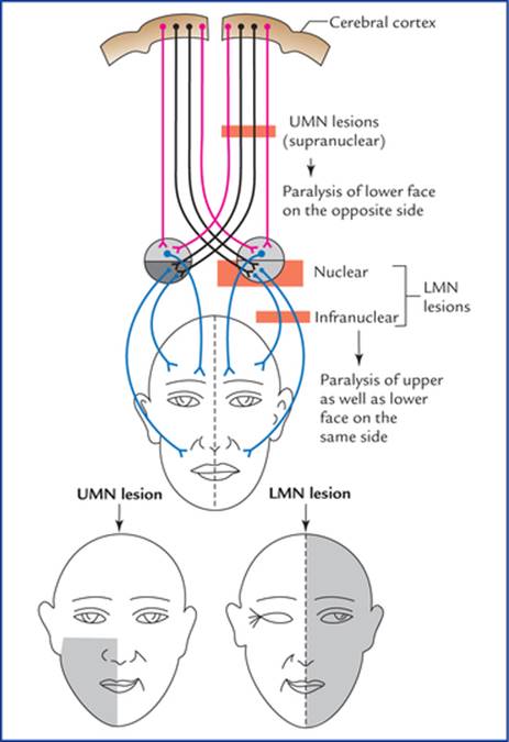
- Characterized by sudden, unilateral facial paralysis due to dysfunction of the facial nerve.
- Symptoms include inability to close the eye, drooping of the mouth, and loss of taste on the anterior two-thirds of the tongue.
- Often idiopathic, but can be associated with infections, trauma, or tumours.
- Facial Nerve Palsy:
- Can result from trauma, infections, tumours, or neurological disorders.
- Symptoms vary depending on the location of the lesion along the nerve's course.
- Ramsay Hunt Syndrome:
- Caused by reactivation of the varicella-zoster virus in the geniculate ganglion.
- Characterized by facial paralysis, ear pain, and vesicles in the ear canal or on the tongue.
Diagnostic Evaluation
- Neurological Examination:
- Assessment of facial symmetry, muscle strength, and taste sensation.
- Evaluation of eye closure, smile, and forehead movement.
- Imaging:
- MRI or CT scans to identify structural abnormalities affecting the facial nerve.
- Electrophysiological Tests:
- Electromyography (EMG) and nerve conduction studies to assess nerve function and muscle activity.
Summary
The facial nerve (cranial nerve VII) is a mixed nerve responsible for motor control of the muscles of facial expression, taste sensation from the anterior two-thirds of the tongue, and parasympathetic innervation of several glands. It originates in the pons, travels through the internal acoustic meatus and facial canal, and exits the skull through the stylomastoid foramen. It has several intracranial and extracranial branches that serve various functions. Clinical conditions affecting the facial nerve include Bell's palsy, facial nerve palsy, and Ramsay Hunt syndrome, which can be diagnosed through neurological examinations, imaging, and electrophysiological tests.

