Related Subjects:
|Osteomalacia-Rickets-Vitamin D
About
- Fractures are seen in 20% of Children who have had an injury
- Chances of fracture up to age of 16 Boys 42% and Girls 27%
- Distal radius, Hand, Elbow, Clavicle, Radius, Tibia
Aetiology
- Epiphysis is the point of bone elongation and are fused in adults
- Children's bones can absorb more energy than adult before fracturing
- Bone in children has good vascular supply so heal well and more quickly
- Bones tend to BOW rather than BREAK
- Sporting accidents, falls from heights, and bike and car accidents.
- May be compounded by poor nutrition and low Calcium and Vitamin D
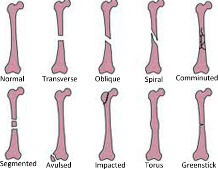
Types of Fractures in children
- Single non displaced
- Transverse
- Oblique
- Spiral
- Comminuted: multiple pieces
- Segmented
- Avulsed: there is a chip of bone hanging off
- Impacted
- Torus: buckles and the bone is dented
- Greenstick: only one side breaks
- Open fracture: means the skin is broken

Fractures Seen
- Clavicle: Most occur in the middle third of the bone 80%. Generally, FOOSH, fall on shoulder, direct trauma
- Humerus: thick bone so less likely to break. Midshaft fractures are rare in kids. Generally caused by a fall. More likely to see fracture round an elbow
- Elbow: 10% of all fractures in children. Most are supracondylar fractures. Ensure not a pulled elbow. Immobilize the elbow before radiographs to avoid further injury from sharp fragments. Flexion 20-30 degrees = least nerve tension
- Radius/Ulna Fracture: Childhood forearm fractures very common following a fall onto an outstretched hand. Young children likely to have sustained greater injuries
- Distal Radius. Peak injury-time coincides with peak growth time. Boys 13-14. Girls 11-12. Most injuries result from FOOSH
- Wrist Fractures. 8 small carpal bones in the wrist. Rare in children under 12 years. Scaphoid fractures most common wrist fractures in adolescents
- Hand injuries: Occur in small bones fingers (phalanges) long bones (metacarpals). Caused from a twisting injury, a fall, a crush injury, or direct contact in sports
- Tibia/Fibula: Tibia and fibula fractures often occur together. Mechanism: falls and twisting injury of the foot
- Toddler's fracture: Children younger than 3 years old learning to walk. No specific injury notable most of the time. Child refuses to weight bear on affected leg
- Ankle Injuries: Typically occur during sports or vigorous play. Sports involving lateral motion and jumping like basketball =higher risk for ankle injuries. Inversion & Eversion injuries
- Foot Injuries. Most foot fractures in kids are minor. Calcaneal fractures -after fall Tarsal fractures? X-Ray. Site of pain-point tenderness
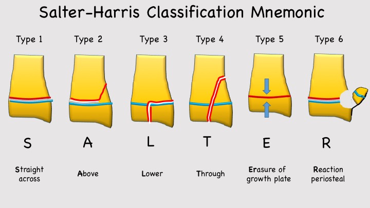
Fractures through Epiphysis: Salter and Harris
- Salter-Harris fractures are often the result of sports-related injuries however they have also been attributed to child abuse, genetics, injury from extreme cold, radiation and medications, neurological disorders, and metabolic diseases which all affect the growth plate
- Type I is a fracture through the growth plate. The fracture line extends through the physis or within the growth plate. Type I fractures are due to the longitudinal force applied through the physis which splits the epiphysis from the metaphysis.
- Type II extends through the metaphysis and the growth plate. There is no involvement of the epiphysis. This is the most common of the Salter-Harris fractures.
- Type III is an intra-articular fracture through the growth plate and the epiphysis. This is rare and when it does occur, it is usually at the distal end of the tibia. If the fracture extends the complete length of the physis, this type of fracture may form two epiphyseal segments. Since the epiphysis is involved, damage to the articular cartilage may occur. One example of this is a Tillaux fracture of the ankle, which is a fracture of the anterolateral aspect of the growth plate and epiphysis[2].
- Type IV extends through the epiphysis, the growth plate and the metaphysis.
- Type V is a crushing type or compression injury of the growth plate injury that affects the growth plate.[3] The force is transmitted through the epiphysis and physis, potentially resulting in disruption of the germinal matrix, hypertrophic region, and vascular supply. Though Harris-Salter V fractures are very rare, they may be seen in cases of electric shock, frostbite, and irradiation. As this fracture pattern tends to result from severe injury, these typically have a poor prognosis leading to bone growth arrest
- There are Type VI-Type IX fractures also but these are rare.

Clinical
- Take a close history. Always consider Non accidental injury
- Get a good history of Trauma?
- Look: Swollen, Red, deformity, open fracture
- Compared to other side
- Check limbs above and below
Check site of tenderness
- Check no neural or vascular injury
- Movement try active and passive movement
- Does the story fit the injury
Investigations
- Calcium and Vitamin D levels if Osteomalacia suspected
- Radiology: ensure you x-ray the correct site. The forearm is not the wrist and the wrist is not the hand. State the precise anatomical part to be examined radiographically. Do not use an X-Ray as a substitution for a proper & thorough examination
Management
- Children heal more quickly than adults, due to a more active periosteum. Rapid healing can sometimes result in re-fracturing. Generally, the management of paediatric fractures is generally quite straightforward and will just require a cast to be applied for a few weeks.
- However, if it is a bad fracture a child may require plates and screws to be put in to align the bone. If the growth plate is damaged, the management can be difficult and can be complicated by progressive deformities with growth as the child gets older. In this instance, a child may require invasive surgery such as leg lengthening.
Fractures through the epiphysis
- Salter-Harris I and II: closed reduction and casting or splinting.
- Salter-Harris III and IV: open reduction and internal fixation (avoiding crossing the physis).
- Salter V fracture: often the diagnosis is not made at the initial presentation. An emergent orthopaedic consultation should be obtained if the fracture is recognized. As these fractures involve the germinal matrix, they have a potential for growth arrest.
- All need a re-examination in seven to ten days is necessary to monitor proper reduction and healing.
- This is also important to determine whether any complications, such as growth arrest, have occurred. If clinically indicated, an additional follow-up radiograph at six and 12 months may be obtained to reassess for any growth arrest.
- The complications include growth arrest with potential for deformity and limb length discrepancy. Entrapment of the periosteum within the fracture is a rare complication that requires an MRI scan. Beware that entrapped periosteum can prevent a complete reduction of the fracture.
References

