Overview of the Midbrain
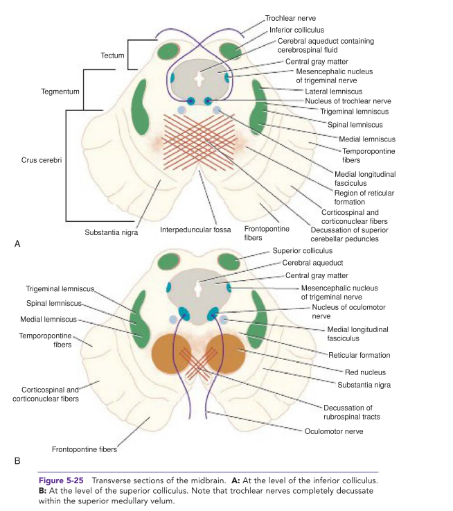
The midbrain, or mesencephalon, is the uppermost part of the brainstem, located between the diencephalon and the pons. It plays a critical role in vision, hearing, motor control, sleep/wake cycles, arousal, and temperature regulation. The midbrain contains several important nuclei and structures essential for these functions.
Anatomy of the Midbrain
- Structural Divisions:
- Tectum:
- The dorsal part of the midbrain, containing the superior and inferior colliculi.
- Superior Colliculi: Involved in visual processing and eye movements.
- Inferior Colliculi: Involved in auditory processing.
- Tegmentum:
- The central part of the midbrain, containing the red nucleus, substantia nigra, and various cranial nerve nuclei.
- Cerebral Peduncles:
- The ventral part of the midbrain, containing the large bundles of fibers (corticospinal, corticobulbar, and corticopontine tracts) that travel between the cerebrum and the spinal cord or brainstem.
Nuclei in the Midbrain
- Cranial Nerve Nuclei:
- Oculomotor Nerve (CN III) Nucleus:
- Controls most of the eye's movements, including constriction of the pupil and maintaining an open eyelid.
- Trochlear Nerve (CN IV) Nucleus:
- Controls the superior oblique muscle of the eye, which is responsible for downward and lateral movement.
- Edinger-Westphal Nucleus:
- Provides parasympathetic input to the eye, controlling the constriction of the pupil and accommodation of the lens.
- Red Nucleus:
- Involved in motor coordination, particularly of the upper limbs.
- Receives input from the cerebellum and motor cortex and projects to the spinal cord.
- Substantia Nigra:
- Part of the basal ganglia, it plays a critical role in reward, addiction, and movement.
- Divided into two parts:
- Pars Compacta: Contains dopaminergic neurons that project to the striatum, important for regulating movement.
- Pars Reticulata: GABAergic neurons that project to various thalamic and brainstem areas.
- Periaqueductal Gray (PAG):
- Surrounds the cerebral aqueduct and is involved in pain modulation and defensive behaviors.
- Plays a role in the descending control of pain, interacting with the opioid system.
Functions of the Midbrain
- Visual and Auditory Processing:
- The superior colliculi coordinate visual reflexes and eye movements.
- The inferior colliculi process auditory information and are involved in auditory reflexes.
- Motor Control:
- The red nucleus and substantia nigra are essential for the coordination and regulation of voluntary movements.
- Pain Modulation:
- The periaqueductal gray modulates pain perception by interacting with the descending pain control pathways and the opioid system.
- Autonomic Functions:
- The Edinger-Westphal nucleus controls the pupillary light reflex and lens accommodation.
- Sleep and Wakefulness:
- The midbrain, through its connections with the reticular formation, plays a role in maintaining arousal and regulating the sleep-wake cycle.
Clinical Relevance
- Parkinson's Disease:
- Characterized by the degeneration of dopaminergic neurons in the substantia nigra pars compacta, leading to symptoms such as bradykinesia, tremors, and rigidity.
- Midbrain Lesions:
- Can result from strokes, tumours, or trauma, leading to deficits in motor control, eye movements, and sensory processing.
- Example: Weber's syndrome, characterized by ipsilateral oculomotor nerve palsy and contralateral hemiparesis due to a lesion in the midbrain.
- Vertical Gaze Palsy:
- Lesions affecting the dorsal midbrain, particularly the region around the superior colliculi, can impair the ability to move the eyes vertically.
Summary
The midbrain is a crucial structure within the brainstem that integrates sensory and motor pathways, modulates pain, and regulates autonomic functions. Its nuclei, such as the substantia nigra, red nucleus, and cranial nerve nuclei, play essential roles in movement, vision, hearing, and arousal. Understanding the anatomy and functions of the midbrain is vital for diagnosing and managing neurological conditions that impact this central area of the brain.

